No products in the cart.
No products in the cart.
No products in the cart.
No products in the cart.
Home » Neurological Recovery Blog » Stroke » Surgery for Stroke: Hemorrhagic vs. Ischemic Interventions
Last updated on March 4, 2024

A stroke can be an incredibly scary event, sometimes requiring emergency surgery. While stroke surgery is not uncommon, the many unknowns can be overwhelming for both survivors and their loved ones. The specific surgery you may receive depends on the type of stroke you experienced and the location of the stroke within the brain. Following stroke surgery, you will then begin a personalized stroke rehabilitation program to help you regain your function and independence.
Not all cases of stroke require surgery. However, we hope to help you better understand the different stroke surgery interventions you may encounter. To begin, we will review the two main types of stroke. Then we will discuss some of the surgical options that help contain or stop tissue damage in the brain. Finally, we will explain your next steps in the recovery process after surgery.
Feel free to use the following jump links to help you navigate this article:
Broadly, a stroke refers to the interruption of the brain’s normal blood flow. This interruption leads to cell death and tissue damage in the brain. There are two main types of stroke, known as hemorrhagic stroke and ischemic stroke. Both types of stroke are very different, and they are treated differently, too.
A hemorrhagic stroke takes place when an artery in the brain bursts or ruptures. Most commonly, hemorrhagic strokes are caused by hypertension, or high blood pressure. This type of stroke often affects the brain’s basal ganglia, although many other areas of the brain can be affected by a hemorrhagic stroke. Hemorrhagic stroke causes bleeding within the brain and, consequently, increases pressure and swelling. This bleeding is referred to as an intracerebral hemorrhage which can lead to a hematoma.
An ischemic stroke is caused by the blockage of an artery in the brain. This causes the area of the brain fed by that artery to lose its blood supply. As a result, brain cells do not receive the oxygen-rich blood needed to function and tissue damage takes place. Around 87% of strokes are ischemic and are caused by a blood clot traveling from another place in the body. Commonly, these clots (or embolisms) originate in the heart and float to the brain. While different arteries can entrap a clot, the most common are the middle cerebral artery (MCA) and the anterior cerebral artery (ACA).
Early stroke detection is very important and can help reduce tissue damage in the brain. Familiarizing yourself with the different stroke warning signs can help you or a loved one access timely stroke treatment. These stroke signs can include drooping of the face, weakness of an arm on one side of the body, slurred speech, or a sudden severe headache. If you or a loved one demonstrate these signs, get emergency help immediately.
If you are experiencing a stroke, upon arrival to the hospital, your medical team will work to determine which type of stroke you are experiencing. This diagnosis process will likely involve a variety of tests and different types of imaging. Once the type and location of the stroke are determined, surgery may be necessary.
Stroke surgery for a hemorrhagic stroke focuses on 3 goals. These goals are stopping the bleeding, resolving the hematoma (collection of clotting blood outside the blood vessels), and relieving intracranial pressure (pressure within the skull). In some cases, these goals can be achieved with conservative interventions. Using medication or other therapies, hematomas can sometimes be resolved without surgical intervention. However, increased pressure on the brain and impaired blood flow may dictate the need for surgery after a hemorrhagic stroke.
Depending upon the stroke severity and the patient’s condition, surgery is often performed within the first 48 to 72 hours. However, sometimes doctors must wait longer if the patient’s condition needs to stabilize before operating. To help you understand what you may encounter, let’s review some common types of stroke surgery used to treat hemorrhagic stroke.
Stroke surgery for an ischemic stroke is generally less invasive. The blockage responsible for an ischemic stroke restricts blood flow to the brain. The longer this is left untreated, the more damage may take place to brain tissue. This can increase the severity of the stroke and lead to more extensive secondary effects.
Ischemic strokes can be treated with clot-busting drugs like tissue plasminogen activator (tPA) under the appropriate circumstances. TPA can be administered within 3-4 hours of symptom onset. Once administered, tPA helps dissolve the clot and restore blood flow to the brain. In some cases of ischemic stroke, however, surgery may be necessary to remove the clot.
The stroke surgery most frequently used to treat an ischemic stroke is a mechanical embolectomy or thrombectomy. During this procedure, the blood clot in the brain is removed using a specialized clot-removal device. This device is inserted into an artery via a catheter in order to reach the clot. This special catheter is generally inserted in the groin, making this procedure much less invasive than brain surgery. Once the clot is secured, the device is then withdrawn and the clot is removed along with it.
To help you visualize this procedure, here is a video that shows how a mechanical embolectomy or thrombectomy works:
Recovery after an intensive stroke surgery like a craniotomy generally takes 6-8 weeks. During this time, it is important to follow the guidelines provided to you by your neurosurgeon. This will include information on the care of your incision, precautions for your daily activities, and warning signs of infection or hemorrhage. Additionally, you will be followed closely by medical staff to ensure you do not experience additional complications.
Following other surgeries, the initial post-operative recovery period may be shorter. For example, the best possible outcome after a mechanical embolectomy surgery of ischemic stroke may include an individual up and walking within days. Regardless of which surgery you have, it is vital to understand and follow instructions from your healthcare team to maximize your outcome.
Once you are medically stable, it will be crucial to start rehabilitation to help you regain any functions lost due to the stroke. Stroke recovery treatments include physical therapy, occupational therapy, and speech therapy. Whether you participate in intensive inpatient stroke rehab after surgery or are discharged to home, it is important to stay consistent with your exercise program. This will provide your brain with the stimulation it needs to make the greatest recovery and increase your independence.
Stroke surgery may be needed to restore normal blood flow in the brain. While less-invasive treatments can be used, in some cases, surgery is necessary to help stop tissue damage in the brain and save the life of the individual. The type of stroke surgery required is determined by the type, severity, and location of the stroke. For example, the two main types of stroke (hemorrhagic and ischemic) require different types of surgery to restore normal blood flow in the brain.
If the stroke is caused by a burst artery (hemorrhagic stroke), neurosurgeons may perform a craniotomy to open up the skull and relieve intracranial pressure. Additionally, less invasive procedures like external ventricular drainage or stereotactic aspiration can be used to drain a hematoma.
When the stroke is caused by a blood clot (ischemic stroke) and cannot be treated with drugs, surgery still may be necessary. A mechanical embolectomy or thrombectomy may be performed to remove the clot via a catheter and clot-removal device. By removing the clot, normal blood flow to the brain can be restored.
Each type of surgery comes with risks and rewards, and it’s best to discuss your unique circumstances with your medical team. However, because stroke is a medical emergency, some decisions are made in high-pressure situations with little time to discuss the details considered for the decisions the medical professionals must make.
When a stroke happens, you often must trust that your medical team will make the best possible decision for you or your loved one. However, the more informed you are, the easier it is to understand the rationale for every decision that is made. Additionally, the more informed you can be, the more helpful you are to the medical team in their decision process. No two medical cases are exactly alike.
While navigating life after stroke can feel overwhelming, there are many tools available to help you on your journey. Once you are medically stable, your rehabilitation process will begin. Stay consistent with your rehab program and keep working to reach your unique post-stroke goals after surgery.

Get our free stroke recovery ebook by signing up below! It contains 15 tips every stroke survivor and caregiver must know. You’ll also receive our weekly Monday newsletter that contains 5 articles on stroke recovery. We will never sell your email address, and we never spam. That we promise.


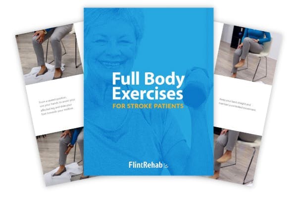
Do you have these 25 pages of rehab exercises?
Get a free copy of our ebook Full Body Exercises for Stroke Patients. Click here to get instant access.
“My name is Monica Davis but the person who is using the FitMi is my husband, Jerry. I first came across FitMi on Facebook. I pondered it for nearly a year. In that time, he had PT, OT and Speech therapy, as well as vision therapy.
I got a little more serious about ordering the FitMi when that all ended 7 months after his stroke. I wish I hadn’t waited to order it. He enjoys it and it is quite a workout!
He loves it when he levels up and gets WOO HOOs! It is a wonderful product! His stroke has affected his left side. Quick medical attention, therapy and FitMi have helped him tremendously!”
FitMi is like your own personal therapist encouraging you to accomplish the high repetition of exercise needed to improve.
When you beat your high score or unlock a new exercise, FitMi provides a little “woo hoo!” as auditory feedback. It’s oddly satisfying and helps motivate you to keep up the great work.
In Jerry’s photo below, you can see him with the FitMi pucks below his feet for one of the leg exercises:
Many therapists recommend using FitMi at home between outpatient therapy visits and they are amazed by how much faster patients improve when using it.
It’s no surprise why over 14,000 OTs voted for FitMi as “Best of Show” at the annual AOTA conference; and why the #1 rehabilitation hospital in America, Shirley Ryan Ability Lab, uses FitMi with their patients.
This award-winning home therapy device is the perfect way to continue recovery from home. Read more stories and reviews by clicking the button below:
Grab a free rehab exercise ebook!
Sign up to receive a free PDF ebook with recovery exercises for stroke, traumatic brain injury, or spinal cord injury below: