The effects of a stroke vary widely, but one common question from survivors is “How does a stroke affect the muscular system?” There are many ways muscle function after stroke can be affected, which can cause survivors to feel overwhelmed and confused by their unique symptoms. However, there is hope for improvement in muscle function through dedicated rehabilitation.
In this article, we will review some of the most common ways a stroke can affect the muscular system and the reasons behind these changes. Then, we will discuss how survivors can recover muscle function after stroke.
Feel free to use the links below to jump straight to any specific section:
How Does a Stroke Affect the Muscular System?
The brain controls both involuntary and voluntary muscle activity by sending neural messages to the muscles. Many different areas of the brain contribute to the production and coordination of muscle function to make your movements smooth and precise. That being said, the messages to initiate movement primarily originate in the motor cortex, which is found in the brain’s frontal lobe.
The motor cortex transmits these signals to the spinal cord, where they can reach the motor neurons. When these neurons fire, the impulse travels to the specific muscle, telling the muscle to contract or relax. When a stroke occurs, however, this complex process can be disrupted.
A stroke takes place when the blood supply to the brain is compromised, either due to a blocked artery or a burst artery. Depending on the severity and location of the stroke, the resulting tissue damage in the brain often leads to changes in how the brain and muscles communicate. In the following sections, we will discuss the most common ways that a stroke can affect the muscular system in more detail.
Changes in Muscle Function After Stroke
A stroke can negatively impact muscle function, tone, and coordination. This can extend to a variety of muscle groups and these effects can range from mild to severe. Since we know that dedicated rehabilitation can help individuals recover movement, it is helpful to understand which movement dysfunction you are experiencing. The following are a few common examples of changes in muscle function after a stroke:
1. Hypotonia (Low Muscle Tone)
Hypotonia, or flaccidity, refers to low or decreased muscle tone. Normally, muscles will maintain a certain level of contraction even when they’re relaxed, which is what makes up normal muscle tone. This resting tone allows you to do things like sit or stand up straight without thinking about it. However, with hypotonia, the muscles do not have this healthy amount of contraction and may feel flaccid or “floppy”.
Hypotonia is a separate condition from muscle weakness, although the two can co-exist, which is common after stroke. In the case of stroke, hypotonia is caused directly by the tissue damage that has occurred in the brain. Hypotonia is a major contributor to other complications like shoulder subluxation after stroke.
2. Hypertonia (High Muscle Tone)
Hypertonia refers to a high amount of muscle tone and increased tension in the muscles. This excess muscle tone occurs when a stroke damages the part of the brain that sends inhibitory signals to the muscles. These signals instruct the muscles to relax when necessary but can be interrupted as a result of the stroke. Consequently, the muscles continuously fire, causing increased muscle tension or stiffness.
Hypertonia typically affects the limbs after stroke, most commonly impacting the arm and hand. If hypertonia affects a patient’s arm, for example, it can cause the arm to feel stiff and difficult to move. It also commonly affects the hand, causing the affected hand to stay clenched into a fist as the fingers may be difficult to move.
If hypertonia persists for too long without intervention, contractures can develop. Contractures occur when muscle, tendon, ligament or skin fibers shorten and become stiff. This contraction of the fibers causes a decrease in the affected joint’s range of motion and a restriction of function. While contractures can affect any joint, the most common include the hips, knees, ankles, elbows, wrists, and shoulders. Contractures typically occur in the areas where hypertonia is most severe and can also be impacted by spasticity, which we will review next.
3. Spasticity
Spasticity is a more specific type of hypertonia that often appears after a stroke. This secondary effect is velocity-dependent and is associated with increased activity of the muscle stretch reflex. The muscle stretch reflex (myotatic reflex) is a contraction that occurs in response to stretching within the muscle.
When you stretch a muscle, its nerve activity increases. Normally, the increased activity triggers a contraction in the muscle fibers to lightly resist the stretching that is taking place. This prevents muscles from stretching too far and tearing, working as a protective mechanism to avoid injury.
This stretch reflex is controlled by the central nervous system. Normally, the muscles and the brain communicate with each other in a constant exchange of information. This allows the brain to know when to contract the muscles and when to release them. However, after a stroke, this communication is often disrupted, which leads to an imbalance of signals in the muscles.
As a result of interrupted communication, the muscle stretch reflex may be overactive. This can cause the muscles to contract or spasm involuntarily. These spasms are worsened by movement, which is what distinguishes spasticity from general hypertonia. Generally, the faster the movement, the more intense the spasm or contraction will be. Spasticity affects around 38% of survivors after their first stroke, which can present an additional challenge to everyday movements and tasks.
4. Hemiplegia and Hemiparesis
A common way that a stroke affects the muscular system is by causing hemiplegia or hemiparesis. Both conditions result from tissue damage within the brain that interrupts communication with the muscles. Specifically, these terms refer to paralysis of one side of the body (hemiplegia) or weakness of one side of the body (hemiparesis).
Each hemisphere (half) of the brain controls movement on the opposite side of the body. When a stroke affects one side of the brain, paralysis or weakness will generally present on the other side of the body, opposite to where the stroke occurred. For example, if a survivor has experienced a stroke in the left side of their brain, they may present with right hemiplegia.
When a stroke impacts the areas of the brain that control muscle movement, the signals between the brain and the muscles can become weakened or lost. As a result, the muscles are not able to respond as well to the brain’s directions, and paralysis or weakness can set in. This is not because of damage to the muscles themselves, but rather the interruption of the neural signals from the brain to those muscles.
When these neural signals are severed completely, this results in paralysis after stroke, or hemiplegia. However, if the connection is damaged but not lost completely, some neural signals can still pass to the muscles. As a result, muscle activation may be significantly weaker, but still possible. This is considered hemiparesis, which is less severe than hemiplegia.
5. Muscle Atrophy
Muscle atrophy is a progressive loss of muscle mass and strength that can take place after a stroke. This deterioration of muscle tissue occurs when a stroke survivor’s muscles are not being used regularly. This is common after a stroke since muscle weakness or paralysis may prevent survivors from moving certain muscles for extended periods. One recent study found that the prevalence of sarcopenia, or muscle loss, increased from 15% pre-stroke to around 50% just one month after stroke.
Aside from the direct weakness that occurs due to inhibited muscle function, there are other reasons why you might experience muscle atrophy. To help you better understand, let’s review three of the most common causes of atrophy after stroke.
Additional Causes of Muscle Atrophy
- Learned nonuse. As we’ve discussed already, stroke often causes weakness or paralysis on one side of the body. This weakness can prompt you to solely rely on your unaffected side to perform tasks. While this may be more efficient, this contributes to learned nonuse. When your affected side is not used at all, your brain can lose its connection to those muscles. This nonuse will cause muscles to atrophy and is especially prevalent in survivors affected by left-side neglect.
- Prolonged hospitalization. In cases of severe stroke, survivors may need to remain in the hospital for several weeks to become medically stable before transitioning to rehab. This prolonged inactivity can cause muscles to rapidly lose strength and mass. While consistent rehabilitation can help survivors gain back muscle function, this process takes time and dedication.
- Malnutrition. Depending on the area of the brain affected, stroke can cause a survivor to have difficulty chewing and swallowing food. This condition, known as dysphagia, affects 30-50% of stroke survivors and can lead to a variety of complications including aspiration pneumonia and malnutrition. Malnutrition can speed up the process of muscle atrophy, especially when combined with inactivity.
As you can see, many stroke-related effects can lead to muscle loss. Therefore, to reverse atrophy and improve muscle function after stroke you will need to address these underlying causes. This can happen when you work closely with your healthcare team to identify your symptoms and create a plan that meets your unique needs.
Restoring Muscle Function After Stroke Through Neuroplasticity
Now that we have answered the question of how does a stroke affect the muscular system, let’s discuss the brain’s natural process of overcoming the effects of a stroke. This process, called neuroplasticity, is the foundation for a stroke survivor’s rehabilitation journey. Neuroplasticity refers to the brain’s ability to reorganize nerve cells and form new neural pathways. These new pathways allow healthy, undamaged portions of the brain to take over control from areas affected by the stroke.
To maximize neuroplasticity, you must engage in consistent therapeutic exercise. The more you practice an activity, such as moving your arm, the more it reinforces new neural pathways that control that function. The more you strengthen those pathways, the stronger the connection between your brain and muscles will become.
However, this poses a dilemma for stroke survivors with hemiplegia or other changes in muscle function. Fortunately, there are many ways to activate neuroplasticity, even if you are unable to perform active movement with your affected limbs.
The Benefits of Passive Range-of-Motion Exercise
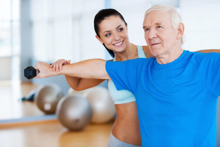
Passive range-of-motion exercises are an effective way to activate neuroplasticity when it is difficult to move on your own. With this technique, a therapist or caregiver can help move your affected limbs through their full range. Additionally, you can perform a passive exercise independently by using your unaffected limbs to move your affected ones. As simple as this sounds, it will significantly aid your stroke recovery by sparking neuroplasticity.
Recent research shows that passive movement activates the same parts of the brain that active movement does. This is especially true when you are watching the movements and paying close attention to the treatment. By triggering neuroplasticity, passive exercises can help reestablish communication with your brain and muscles and rewire neural connections for movement.
Over time, passive exercise can help increase muscle function after stroke and reduce paralysis and spasticity. In addition, performing passive range-of-motion exercises will allow you to maintain mobility, helping to prevent contractures. This is critical for preventing further loss of muscle function. Eventually, you may even recover enough strength to move independently.
The Importance of Active Exercise at Home
The more you engage in passive exercise, the stronger the connections between your brain and muscles can become. In some cases, the strengthening of these neural pathways can allow you to regain muscle activation in your affected limbs. Once you achieve muscle activation in your affected limbs, you must participate in repetitive active exercises to continue strengthening those neural pathways.
Active exercises are exercises that require your own muscle contraction to perform the motion, which means you are doing the exercise without assistance. Transitioning from passive to active exercises will allow you to make the most of the changes in function you are experiencing, although this takes high repetition or massed practice. As you practice these exercises, you are promoting substantial changes in your brain, which can help you regain further function and increase your independence.
Ask your therapy team for exercise guidelines and a personalized home exercise program to help kickstart your recovery. They can help you make the transition from active to passive exercises when you are ready and can adjust your program based on your progress.
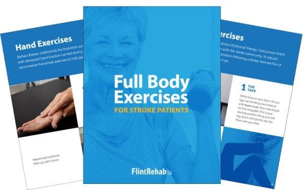
Since high repetition and consistency are key for making gains in muscle function after a stroke, it is important to find ways to stay motivated. One way is to incorporate therapy devices like Flint Rehab’s FitMi. This device can help you accomplish the high repetition necessary to see improvement. In fact, FitMi helped a stroke survivor with arm paralysis experience twitches in his affected arm after three weeks of daily FitMi use.
Understanding “How Does a Stroke Affect the Muscular System?”
Stroke survivors can face a wide range of challenges during recovery and it can be difficult to predict how a stroke might affect muscle function. In addition to hemiplegia or hemiparesis, survivors may be affected by spasticity, muscle atrophy, and even dysphagia. This can have a major impact on daily life, performance of the activities of daily living, and overall independence.
The specific ways a stroke will affect muscle function depend heavily on the stroke’s severity and location. Additionally, the time spent in the hospital during the acute stage of recovery can impact a survivor’s muscle function after stroke. Fortunately, thanks to the brain’s natural neuroplasticity, it is possible to regain some muscle activation and strength.
The key to recovery is engaging in regular and intensive therapy. High repetition of therapeutic exercises will boost neuroplasticity and allow you to see the greatest improvements in function. Even if you experience hemiplegia, passive range-of-motion exercises can help activate the brain and promote recovery. Talk with your rehab team about your options and continue pursuing your goals one step at a time.