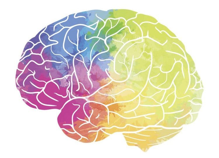
All areas of the brain can be affected by stroke, and every stroke will be accompanied by unique effects. This is because each area of the brain controls different functions, and everyone’s brain is wired a bit differently. Therefore, therapists and doctors frequently say, “Every stroke is different, so every recovery will be different.”
In this article, we will discuss 9 major areas of the brain that are commonly affected by stroke. Although the effects of a stroke vary from person to person, we will review some common secondary effects of a stroke in each of these areas. While each case will be unique, this can provide a rough idea of what to expect from each type of stroke and tips for navigating the recovery journey.
Feel free to use the links below to jump straight to any section:
- Frontal lobe stroke
- Parietal lobe stroke
- Temporal lobe stroke
- Occipital lobe stroke
- Brain stem stroke
- Cerebellar stroke
- Thalamic stroke
- Basal ganglia stroke
- Internal capsule stroke
What Is a Stroke?
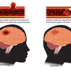
A stroke occurs when the supply of blood in the brain becomes compromised. One way this can happen is by a blood clot obstructing an artery and stopping blood flow to an area of the brain. This is called an ischemic stroke. The other is when an artery in the brain bursts and leads to bleeding inside the brain, called a hemorrhagic stroke.
During a stroke, the affected areas of the brain do not receive enough oxygen-rich blood. Additionally, pooling blood can place increased pressure on the brain. As a result, brain tissue begins to die. Depending on the area of the brain affected by stroke, this damage can cause changes in certain sensory, motor, or cognitive functions. This is why timely emergency care is critical to minimize tissue damage and reduce the severity of the stroke. While some strokes can be treated with medication, others may require surgical intervention.
Although it’s impossible to revive dead brain cells, recovery is possible through neuroplasticity. This process allows healthy parts of the brain to take over the functions of areas damaged by the stroke. While this process occurs naturally and contributes to spontaneous recovery, neuroplasticity can also be optimized through dedicated rehabilitation exercises.
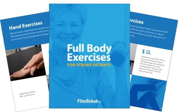
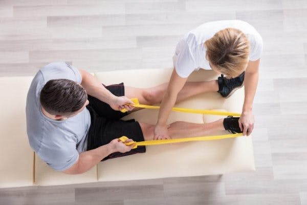
The goal of stroke rehabilitation is to restore or compensate for the secondary effects sustained due to the stroke. These effects vary from person to person based on the size and location of the stroke. By working with your rehab team and staying consistent with your therapy exercises after stroke, you can work to achieve your highest potential and increase your independence.
Cortical Strokes vs. Subcortical Strokes
Before we dive into the different areas of the brain commonly affected by stroke, you should know the difference between cortical and subcortical strokes. This can help you understand what a stroke in the different areas of the brain may entail.
The cerebral cortex or cerebrum is a large part of the brain that includes 4 lobes. These are the frontal lobe, parietal lobe, occipital lobe, and temporal lobe. Strokes in these regions are known as cortical strokes. Aside from the cerebrum, there are subcortical structures that lie deep within the brain. Subcortical refers to the area “below the cortex.” This includes structures such as the thalamus, hypothalamus, and basal ganglia. Strokes in these areas of the brain are known as subcortical strokes.
The arteries that supply the subcortical areas of the brain are smaller and more delicate. As such, a hemorrhagic subcortical stroke can occur when these delicate arteries rupture due to high blood pressure or other complications. However, the subcortical area of the brain can still be affected by ischemic stroke. When an ischemic stroke occurs in the subcortical regions, it’s referred to as a lacunar stroke.
There are many differences between cortical and subcortical strokes. For example, cortical strokes often impact higher-level functioning. Therefore, cortical strokes often result in effects like aphasia or unilateral neglect. On the other hand, subcortical strokes are generally associated with motor symptoms, such as hemiparesis. We will review the specific ways these areas of the brain can be affected by stroke in the next section.
Major Areas of the Brain Affected by Stroke
Below, you’ll learn about 9 different areas of the brain that can be impacted by stroke. In each section, you will find a summary of the effects of each type of stroke to help you understand common patterns. Additionally, you can click the link in each section to learn more information.
Remember, the effects of a stroke will vary from person to person, so it can help to reference a full list of the secondary effects of stroke. This may give you an even better idea of what to expect. Then, you can work to create a recovery plan that meets your specific needs and goals after stroke.
1. Frontal Lobe Stroke
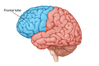
Almost one third of the cerebrum is comprised of the frontal lobe. Therefore, it should be no surprise that the frontal lobe plays a role in many functions. Motor skills, executive functioning, speech, language, and social skills are all controlled in some part by the frontal lobe.
Effects of a frontal lobe stroke (a type of cortical stroke) include motor impairments, problem-solving and judgment issues, behavioral changes, personality changes, and difficulty with speech. These speech difficulties can include aphasia, dysarthria, or apraxia of speech, among others.
Learn more about frontal lobe stroke »
2. Parietal Lobe Stroke
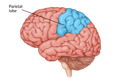
A parietal lobe stroke mostly affects sensory interpretation, language skills, and spatial awareness. Some secondary effects of this cortical stroke include hemineglect and poor proprioception or body awareness. Additionally, survivors of parietal lobe stroke may experience difficulty writing (agraphia), difficulty reading (alexia), or difficulty speaking (aphasia), among other symptoms.
Learn more about parietal lobe stroke »
3. Temporal Lobe Stroke
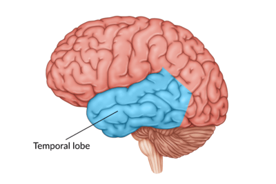
The temporal lobe, also part of the cerebrum, is an area of the brain that controls language comprehension, hearing, and other sensory processes. A temporal lobe stroke may affect hearing, vision, and speech comprehension, along with other secondary effects.
Learn more about temporal lobe stroke »
4. Occipital Lobe Stroke
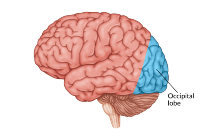
The occipital lobe plays a large role in your vision. As a result, an occipital lobe stroke (a type of cortical stroke) often results in vision difficulties. This can include central vision loss, cortical blindness, visual hallucinations, or other secondary effects like prosopagnosia.
Learn more about occipital lobe stroke »
5. Brain Stem Stroke
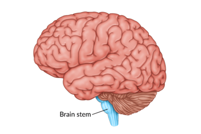
The brain stem is comprised of the midbrain, pons, and medulla oblongata. A stroke in any of these areas is considered a brain stem stroke. While less common than other types of strokes, brain stem strokes are often life-threatening because of this area’s vital functions.
The brain stem controls basic body functions like breathing, sweating, and consciousness. Therefore, common changes caused by a brain stem stroke include coma, difficulty breathing, and difficulty swallowing (dysphagia), among other effects.
Learn more about brain stem stroke »
6. Cerebellar Stroke
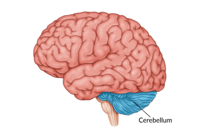
A stroke in the cerebellum is called a cerebellar stroke. The cerebellum controls some sensory functions and voluntary movements, specifically balance and coordination. Effects of a cerebellar stroke may include ataxia, balance difficulties, and spatial awareness issues, among others. Additionally, vertigo commonly accompanies a stroke in the cerebellum.
Learn more about cerebellar stroke »
7. Thalamic Stroke
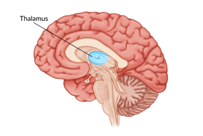
When a stroke affects the thalamus, it’s called a thalamic stroke and is considered subcortical. One of the biggest effects of a thalamic stroke is sensory issues because the thalamus relays 98% of all sensory input. While numbness and sensory impairments are extremely common after a thalamic stroke, other effects like attention and memory changes can also take place. Central post-stroke pain, a type of chronic pain, is also common after a thalamic stroke.
Learn more about thalamic stroke »
8. Basal Ganglia Stroke
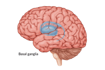
The basal ganglia are most associated with emotions, voluntary muscle control, cognitive function, and memory. Therefore, basal ganglia strokes often result in emotional blunting, post-stroke depression, and motor impairments, among other effects. Additionally, this type of subcortical stroke can cause survivors to experience changes in learning.
Learn more about basal ganglia stroke »
9. Internal Capsule Stroke
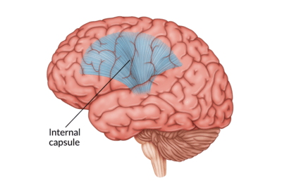
The internal capsule is another subcortical region that lies deep within the brain. This unique area connects different parts of the brain and plays a large role in movement. Therefore, motor impairments are the most common effect of an internal capsule stroke. This can include hemiplegia and facial weakness, although hearing, sensation, and cognition can also be affected by stroke in this area of the brain. If motor impairments are the only secondary effect, then this type of stroke is called a pure motor stroke.
Learn more about internal capsule stroke »
Left Hemisphere vs. Right Hemisphere Stroke
Along with different lobes and structures, the brain is also divided into two halves, called hemispheres. Each hemisphere contains the four cortical lobes, as well as subcortical structures. When reviewing the different areas of the brain that can be affected by stroke, it’s also helpful to look at differences between the two hemispheres.
The two hemispheres of the brain house many of the same functions. However, there are some differences, and these differences vary from person to person. Generally speaking, the left hemisphere houses areas that produce language. This is why language difficulties are often associated with left hemisphere strokes. On the other hand, the right hemisphere is believed to control self-awareness and object recognition.
Furthermore, each hemisphere controls movement on the opposite side of the body. Usually, a left hemisphere stroke will cause motor impairments on the right side of the body. In contrast, a right hemisphere stroke will likely impair movement on the left side of the body and can lead to left hemiplegia or hemiparesis. When stroke impacts both hemispheres, it’s possible to experience motor changes on both sides of the body.
Areas of the Brain Affected by Stroke: Recovery
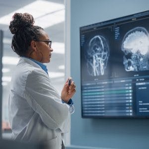
If you are a stroke survivor, it’s important to talk to your neurologist about your specific diagnosis. Ask them about the location and the type of stroke you experienced, as it may help you to identify and understand what secondary effects to expect. Once you understand the location and effects of your stroke, rehabilitation can proceed with more efficiency.
The stroke recovery process is unique to each individual because every stroke is different. The specific symptoms you experience are based largely on the area of your brain affected. However, there is always hope for recovery through a dedicated rehab program. This is just as true for those in the chronic stages of recovery. The most important thing to do is never give up hope during your stroke journey.
Keep pursuing rehabilitation so that you can get as close to a full recovery from stroke as possible. With time, you can see the return of important functions which can increase your independence. Lean on those around you, seek support from your community, and never stop pursuing your recovery goals.